Mathematics as a Bridge from the Laboratory to the Clinic - Developing Mathematical Models

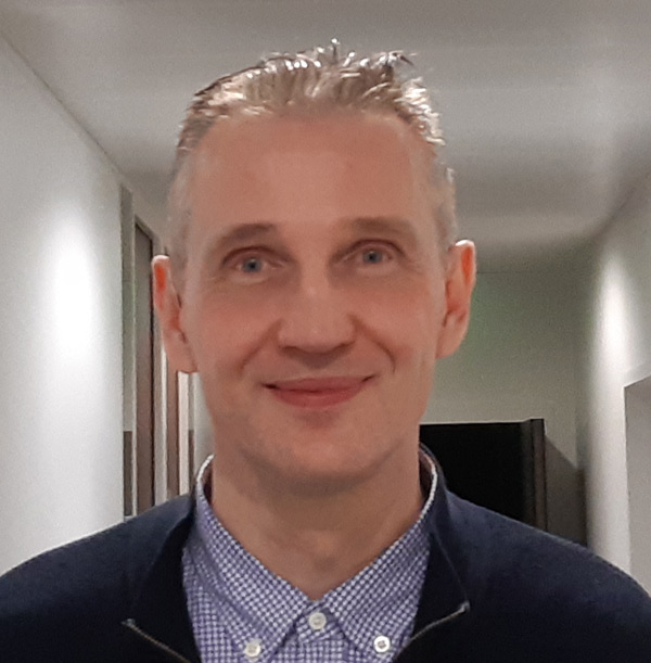
Developing mathematical models of biological systems is very time consuming and challenging for scientists. Yet this is the only way for many experimental results to actually reach patients. The computer scientist Dr. habil. Dirk Drasdo from the systems biology research network LiSyM explains some of the technical issues involved when creating models. A simulations expert, Drasdo uses the liver model he has developed as an example to explain how chemistry, physics, biology, pharmacy, and medicine are transformed into mathematical equations and simulations.
Modern systems biology and systems medicine research is not possible without mathematical models. They are the only way to combine all experimental results from various sources in a meaningful way, including those from a test tube, cell cultures, and living organisms. But how is this achieved in detail, how are the processes of life translated into mathematics? Dr. habil. Dirk Drasdo provides a clear explanation using his liver model as a basis. The development of complex models that closely represent reality is dependent on interdisciplinary, stabile research networks like LiSyM. There is a great deal of effort involved in developing entire models, often even integrated model components. As Drasdo emphasizes, “Thousands of simulations are sometimes necessary for certain steps.”
Reaction equations can be integrated relatively easily
Complex mathematical models consist of countless equations. Reaction equations indicate the changes in the ratios of substances involved in chemical reactions. Such equations can be transformed into mathematical equations relatively easily and can include additional information – for example, whether catalysts were involved in the reactions. This means that biochemical reactions catalyzed by enzymes can also be formulated as mathematical equations. As a result, processes within individual cells, such as intracellular detoxification reactions, can be integrated in the models. Mathematical equations derived from chemical reactions can also reflect intracellular processes, including the exchange of signaling molecules, which occurs between liver cells to coordinate their responses in the regeneration of the liver after a paracetamol poisoning.
Not even all cells of the same type are truly identical
“We solve these equations in every single cell of a simulated tissue,” says Drasdo. This is necessary because not even all cells of the same type are completely identical in the functional subdivisions of the liver: the liver lobules. “These lobules are divided into zones,” explains Drasdo. Simply put, each lobule consists of an outer, periportal zone, an inner, pericentral zone, and sometimes even additional zones. “Depending on the zone, the hepatocytes (liver cells) are equipped with different enzymes,” says the researcher, adding: “The activities of these enzymes can vary, depending on the exact position of the cell as well as other factors.” That is why hepatocytes in a liver lobule sometimes use different metabolic pathways – and why many toxic substances result in characteristic patterns of damage in the liver. “After a poisoning with a high, but not extreme, dose of paracetamol, only pericentral hepatocytes die,” explains Drasdo. This is because they are the only cells in which enzymes in the cytochrome P450 system convert paracetamol into the toxic byproduct N-acetyl-p-benzoquinone imine (NAPQI), which damages hepatocytes. In the periportal hepatocytes, on the other hand, no toxic substances are produced when paracetamol is metabolized.
Chronological series of cross-sectional images show how fast tissue dies
Researchers use antibodies to mark these enzymes and other proteins found in tissues. In cross-sectional images of tissue, these markers often allow researchers to detect differences in the spatial distribution and dynamics of many processes. “We can use chronological series of cross-sectional images of tissue, for example, to measure how many cells grow back and when,” says Drasdo. He can also use such a series of images to quantify the necrosis of the liver after poisoning. “For this, we use algorithmic methods to conduct quantitative analyses of images and video clips created using imaging techniques,” he says. Modern intravital imaging methods even enable researchers to make short videos of certain processes in living tissue, so they can calculate the speed of blood and bile flow, for example.
When changes in the shape of the cell occur, each surface point is assigned its own equation
The simulation of cells and tissues goes far beyond the solving of reaction equations. These equations only make up the more chemical side, while motion and dynamics make up the more physical side. “We also represent tissue spatially in all its architecture and dynamics; this is done realistically – much like the figures and environments in modern computer games. But instead of animations, they are simulations,” says Drasdo. In addition to processes inside the tissue, his model also incorporates the active and passive motion of individual cells – for example, when they become deformed through external factors, or when they grow, divide, and die. In the model, a grid connects all the points on a cell’s surface. If, for example, a cell puts external pressure on a neighboring cell, its surface will be most deformed at the point of contact. This means that the grid is most deformed there as well. Because the deformation continues in surrounding cells, the grid conveys this movement in a weaker form to the points directly adjacent, which in turn convey this in yet a weaker form to their neighboring points, and so forth. “It is necessary to calculate a separate equation of motion for each point on the cell’s surface,” explains Drasdo.
Models as complex as rocket science
Last but not least, Drasdo’s team combines the chemical reaction equations with physical equations of motion. This means they connect molecular processes with changes in cellular forms caused by internal or external influences, as well as with processes at the tissue level. The complexity increases with every new component. “The complexity of a patient’s liver will ultimately be comparable to that of a moon rocket,” says the expert. The current liver model is already able to describe exactly where and how quickly certain processes occur across several spatial and temporal scales. It can also describe how these are connected to the intracellular and intercellular environment. “These processes cannot be represented in fixed diagrams. They are too comprehensive, too dynamic, and too complex,” says Drasdo.
“In the long run, our models will be immensely beneficial!”
Another major advantage of these models – namely, that they can master a high degree of complexity – has one disadvantage. “You need stamina and endurance to build them!” says Drasdo. Every small addition, every new component can be immensely time-consuming. “Sometimes we have to do thousands of simulations with just parts of systems,” he says. This is exactly why the development of models needs solid long-term funding as well as larger collaboration efforts or research networks like LiSyM. The data for Drasdo’s model stem from researchers in many specialized disciplines, such as cell metabolism, mechanical properties of cells, signal transduction, and blood flow. Drasdo believes it is all worth the effort: “In the long run, our models will be immensely beneficial!”
Back to Feature Articles